What is UPJ Obstruction?
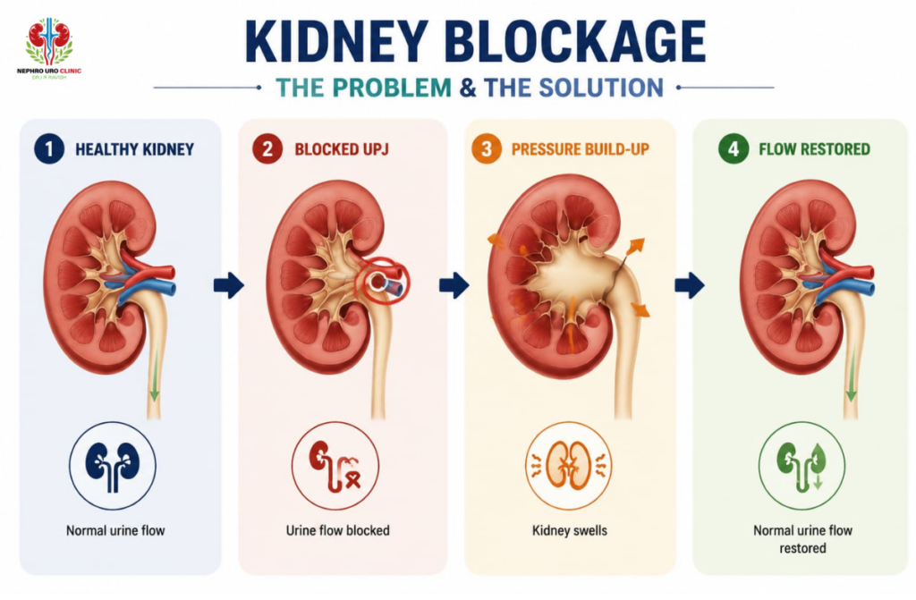
The ureteropelvic junction, or UPJ, is the point where the kidney connects to the ureter. The ureter is the tube that carries urine from the kidney down to the bladder. When this junction becomes narrowed or blocked, urine cannot drain freely from the kidney. This causes urine to back up and collect inside the kidney, a condition called hydronephrosis.
UPJ obstruction can be present from birth (congenital) or develop later in life due to kidney stones, scarring from infection or previous surgery, or external pressure from a blood vessel crossing the ureter at an abnormal angle.
If left untreated, a significant UPJ obstruction gradually damages the kidney by reducing its ability to filter blood. The goal of pyeloplasty surgery in Bengaluru is to remove the blocked segment and restore normal urine flow before permanent kidney damage occurs.
Who Needs Pyeloplasty Surgery?
Not every UPJ obstruction requires immediate surgery. Mild cases in adults may be monitored over time. Surgery is recommended when:
- The obstruction is causing significant pain, typically a dull ache or cramping in the flank or side
- Recurrent urinary tract infections are linked to poor kidney drainage
- Imaging shows that hydronephrosis (swelling of the kidney) is worsening
- Kidney function tests show a decline in the affected kidney
- The patient has kidney stones caused by urine stagnation in the blocked kidney
- In some patients, a poorly draining kidney may contribute to secondary hypertension
In children, UPJ obstruction is one of the most common causes of hydronephrosis detected before or shortly after birth. Early surgical correction in these cases protects kidney development.
Symptoms of UPJ Obstruction
Many patients with mild obstruction have no symptoms at all and are diagnosed incidentally during imaging for another reason. When symptoms are present, they typically include:
- Pain in the side or back, often worse after drinking large amounts of fluid
- Intermittent nausea or vomiting alongside flank pain
- Recurrent urinary tract infections that keep coming back despite treatment
- Blood in the urine
- A palpable lump in the abdomen in infants or young children
- Reduced kidney function detected on blood tests or nuclear scans
How is UPJ Obstruction Diagnosed?
Diagnosis involves a combination of imaging and functional tests.
Ultrasound is usually the first test. It shows the degree of kidney swelling and is safe for all patients including pregnant women and newborns.
CT urogram provides detailed images of the kidney, ureter, and the exact location of the obstruction. It also helps identify crossing vessels that may be contributing to the blockage.
MAG3 or DTPA nuclear scan (renogram) measures how well each kidney is functioning and how effectively it drains urine. This is one of the most important tests for deciding whether surgery is necessary, as it shows the functional contribution of each kidney.
Blood tests including creatinine and eGFR assess overall kidney function.
Types of Pyeloplasty Surgery Available in Bengaluru
There are three main surgical approaches for pyeloplasty. The best option depends on the patient’s age, anatomy, previous surgeries, and the complexity of the obstruction.
Laparoscopic Pyeloplasty
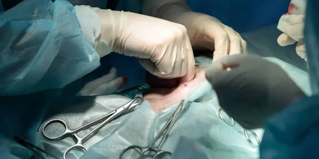
Laparoscopic pyeloplasty is the most commonly performed approach for adults and children generally above the age of two to three years and weighing over 10 to 12 kg, where the abdominal space allows safe instrument placement. It is a minimally invasive procedure using three to four small cuts in the abdomen, each less than 1.5 cm in size. A thin camera called a laparoscope is inserted through one cut, and fine surgical instruments are passed through the others.
The surgeon removes the narrowed or blocked segment of the ureter and reconnects the healthy ureter directly to the kidney’s collecting system. A ureteral stent is placed at the time of surgery to keep the repaired junction open while it heals.
Benefits of laparoscopic pyeloplasty include less postoperative pain, a shorter hospital stay of two to three days, quicker return to normal activity, and minimal visible scarring.
The success rate of laparoscopic pyeloplasty is over 90% in experienced hands and is comparable to open surgery for most patients.
Open Pyeloplasty
Open pyeloplasty uses a larger incision in the flank or abdomen to directly access the kidney and ureter. While it involves a longer recovery than laparoscopic surgery, it remains the preferred option in certain situations.
Open pyeloplasty is recommended for:
- Infants and young children typically under two years of age or below 10 kg, where the anatomy is too small for safe laparoscopic instrument placement
- Patients with complex anatomy or significant scarring from previous surgery
- Cases where a crossing vessel needs to be repositioned alongside the pyeloplasty repair
- Situations where laparoscopic surgery is not feasible for technical reasons
Hospital stay for open pyeloplasty is typically four to five days, and full recovery takes six to eight weeks. The success rate is equivalent to laparoscopic surgery, consistently above 90%.
Robotic-Assisted Laparoscopic Pyeloplasty
Robotic pyeloplasty uses a robotic surgical system to perform the same minimally invasive repair as laparoscopic pyeloplasty, but with enhanced precision. The surgeon controls robotic arms from a console, using instruments with a greater range of motion than the human wrist and a high-definition three-dimensional view of the operative field.
Robotic pyeloplasty is particularly useful for complex reconstructions, revision cases where a previous pyeloplasty has failed, and cases involving significant scarring. Recovery is similar to standard laparoscopic surgery.
The Pyeloplasty Procedure: What Happens Step by Step
Before surgery: You will have blood tests, urine culture, and imaging to confirm the surgical plan. Medications that thin the blood, such as aspirin or warfarin, are usually stopped several days beforehand. You will fast for six to eight hours before the procedure. A urine infection, if present, must be treated with antibiotics before surgery proceeds.
Anaesthesia: Pyeloplasty is performed under general anaesthesia. You will be fully asleep throughout the procedure.
During surgery: The blocked segment of the ureter is identified, excised, and the healthy ureter is carefully reconnected to the kidney’s collecting system. The repair is called a dismembered pyeloplasty, named after the technique of separating and reconstructing the junction. A ureteral stent, often called a DJ stent or double-J stent, is placed to support the repair during healing. In some cases, a drain tube may also be placed near the kidney temporarily.
Duration: Laparoscopic pyeloplasty typically takes two to three hours. Open surgery may take slightly longer for complex cases.
Recovery After Pyeloplasty Surgery in Bengaluru
In the Hospital
Most laparoscopic pyeloplasty patients are discharged within two to three days. Open surgery patients stay four to five days. During this time, pain is managed with medications and urine output is monitored to confirm that drainage has improved.
The Ureteral Stent
After pyeloplasty, a DJ stent remains inside the ureter to keep the repaired junction open while it heals. This stent is temporary. It is removed four to six weeks after surgery in a short outpatient procedure that takes only a few minutes. The removal is done using a thin flexible cystoscope passed through the urethra. A local anaesthetic gel is applied beforehand to minimise discomfort. Most patients find the procedure uncomfortable rather than painful, and can drive home afterward.
While the stent is in place, it is normal to experience urinary frequency, urgency, mild burning during urination, and occasional flank discomfort when passing urine. A small amount of blood in the urine is also common. These symptoms improve once the stent is removed.
When to Call the Clinic After Surgery
Most recovery after pyeloplasty is straightforward, but certain symptoms need prompt attention. Contact Nephro Uro Clinic or go to an emergency room immediately if you notice any of the following:
- Fever above 38.3°C (101°F)
- Severe flank pain or abdominal swelling that is not controlled by prescribed medication
- Complete inability to pass urine or a sudden significant drop in urine output
- Large blood clots in the urine or persistent dark red bleeding that does not settle
- Vomiting that prevents you from keeping fluids or medications down
- Signs of infection at any wound site such as redness, warmth, or discharge
Do not wait to see if these symptoms pass on their own.
At Home
Most patients can return to desk work and light activity within one to two weeks after laparoscopic pyeloplasty. Open surgery patients generally need four to six weeks before returning to normal activity.
Avoid heavy lifting, strenuous exercise, and driving while taking prescription pain medication. Drink two to three litres of water daily to support healing and prevent urinary infections.
Follow-Up
A urine culture is repeated approximately two weeks after surgery to check for infection.
An ultrasound of the kidney is arranged at three months after surgery to confirm that the hydronephrosis is reducing or stable.
A nuclear scan (DTPA or MAG3 renogram) may be done at one year after surgery to assess kidney function and drainage objectively.
Regular follow-up is important. Occasional cases of late re-narrowing can be identified early and managed before kidney function is affected.
Risks of Pyeloplasty Surgery
Pyeloplasty is a safe procedure with a well-established record of success. As with all surgery, there are some risks to be aware of.
Bleeding during or after the procedure is uncommon. Significant bleeding requiring transfusion is rare.
Infection can occur at the surgical site or within the urinary tract. Antibiotics are given around the time of surgery to reduce this risk.
Urine leak from the repair site occurs in a small number of cases and usually resolves with the stent in place and conservative management.
Injury to surrounding structures such as blood vessels or bowel is rare but possible in any abdominal surgery.
Recurrence of obstruction occurs in a small percentage of cases, more often in patients who have had previous pyeloplasty surgery. A revision procedure may be needed in these cases.
Anaesthesia risks are standard for any procedure under general anaesthesia and are discussed by the anaesthetist before surgery.
Success Rate
The success rate of pyeloplasty, whether open or laparoscopic, is consistently reported above 90 to 95% in experienced surgical centres. Most patients experience complete resolution of symptoms, improvement in kidney drainage on post-operative imaging, and stable or improved kidney function on long-term follow-up.
Outcomes are best when surgery is performed by a urologist with specific experience in reconstructive urology and when long-term follow-up imaging is completed.
Pyeloplasty Surgery Cost in Bengaluru
The cost of pyeloplasty surgery in Bengaluru varies depending on the type of surgery chosen (open, laparoscopic, or robotic), the specific clinical requirements of each patient, hospital charges, and post-operative care including stent removal.
For an accurate cost estimate specific to your situation, contact Nephro Uro Clinic directly. A consultation with Dr. I. R. Ravish will include a full assessment of your imaging and test results, a clear recommendation on the surgical approach best suited to you, and a transparent breakdown of expected costs before any decision is made.
When to See a Doctor
If you or your child has been diagnosed with hydronephrosis or UPJ obstruction, or if you are experiencing recurrent flank pain, repeated kidney infections, or have been told your kidney function is declining, an early surgical consultation is advisable.
Earlier treatment preserves more kidney function. Waiting until the kidney is severely damaged reduces the benefit that surgery can provide.
Pyeloplasty Surgery at Nephro Uro Clinic, Bengaluru
Dr. I. R. Ravish is a senior urologist with over 20 years of dedicated urology experience, having completed his MCh in Urology in 2007, specialising in reconstructive and laparoscopic urological surgery. He performs laparoscopic pyeloplasty surgery in Bengaluru for both adult and paediatric patients, and manages complex cases including revision pyeloplasty and obstruction associated with crossing vessels.
Nephro Uro Clinic is located in Jayanagar, Bengaluru, and is accessible to patients from across the city and surrounding areas including Tilaknagar, JP Nagar, Bannerghatta Road, and Electronic City.
To schedule a consultation, contact the clinic directly. Appointments are available for new patients seeking a second opinion, post-diagnosis counselling, and pre-surgical planning.